
The reproducibility crisis in science has been making headlines for years, but there's a quieter version of this problem hiding inside MRI machines - and a new survey paper is shining a spotlight on how researchers are fighting back.
The "Is It the Brain or the Scanner?" Problem
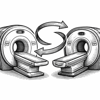
Here's the thing. You'd assume that if you scanned the same brain on two different MRI machines, you'd get the same picture. You would be wrong. Like, impressively wrong.
MRI data collected across different scanners, protocols, or hospitals can look so wildly different that scientists call the discrepancies "batch effects" or "site effects." These aren't subtle variations. They're the kind of noise that can completely drown out the actual biology researchers are trying to study. Imagine trying to compare photos of the same sunset taken on 36 different phones with 36 different Instagram filters. Now imagine your medical diagnosis depends on which phone was used.
A sweeping new review by Qinqin Yang, Firoozeh Shomal-Zadeh, and Ali Gholipour, published in Medical Image Analysis, maps out the entire landscape of methods scientists use to fix this mess (Yang et al., 2026). It's the most comprehensive look yet at MRI harmonization - the art and science of making brain scans play nice with each other.
So What's Actually Going Wrong?
Every MRI scanner is a little bit of a diva. Different manufacturers (Siemens, GE, Philips), different magnetic field strengths, different software versions, different coils - they all introduce their own quirks into the data. It's like every scanner has a slightly different accent, and your analysis pipeline doesn't speak any of them fluently.
This becomes a massive headache for large-scale brain studies. The ENIGMA consortium, which pools data from over 1,400 scientists across 43 countries, has been wrestling with this for over a decade (Thompson et al., 2020). When you're combining brain scans from 200+ institutions, scanner-to-scanner variation isn't a minor footnote - it's a potential study-wrecker.
The Harmonization Toolbox
The review breaks down fixes into a few major categories, and honestly, they read like a tech tree in a strategy game.
Level 1: Fix It Before It Happens. Prospective harmonization means standardizing how scans are acquired in the first place - locking down protocols across sites. Prevention beats cure, but good luck getting 50 hospitals to agree on anything.
Level 2: Fix the Images After the Fact. Retrospective image-level methods use algorithms (increasingly deep learning-based) to make scans from different machines look like they came from the same one. Think of it as Photoshop for brain scans, except the goal is accuracy, not aesthetics.
Level 3: Fix the Numbers. Feature-level methods skip the images and correct the measurements extracted from them. The rockstar here is ComBat, a statistical method with over 8,000 citations that adjusts for scanner-related biases while (hopefully) leaving the biology intact (Fortin et al., 2018). New multivariate extensions like MV-ComBat are pushing this further by harmonizing multiple metrics simultaneously instead of one at a time.
Bonus Level: Traveling Subjects. Some researchers literally send the same people to get scanned at multiple sites. The Oxford-Nottingham Harmonisation resource scanned 20 people on six different scanners across five sites to create a gold-standard reference dataset (Sherwood et al., 2025). It's the scientific equivalent of a taste test, and it's wildly useful.
The Catch (There's Always a Catch)
The review's most important finding is also its most sobering. Current methods are genuinely good at removing scanner-related variability. But here's the unsolved puzzle: how do you prove you didn't accidentally scrub away real biological signal in the process?
Stripping out site effects is one thing. Guaranteeing that the subtle brain differences between a healthy person and someone with early Alzheimer's survived the harmonization pipeline? That's where the field still needs work. The authors argue for standardized validation benchmarks and smarter evaluation strategies - because right now, everyone's grading their own homework.
Why This Matters Beyond the Lab
Large-scale neuroimaging studies are the backbone of modern brain research, from mapping psychiatric disorders to tracking neurodegeneration. A recent multi-center study across 36 datasets showed that harmonization meaningfully boosts the performance of machine learning models trained on brain data (Pomponio et al., 2023). As AI-driven diagnostics inch closer to clinical use, the stakes of getting harmonization right just keep climbing.
Look. Your brain is the most complex object in the known universe. The least we can do is make sure the machines photographing it are speaking the same language.
References
-
Yang, Q., Shomal-Zadeh, F., & Gholipour, A. (2026). Harmonization in magnetic resonance imaging: A survey of acquisition, image-level, and feature-level methods. Medical Image Analysis, 104066. DOI: 10.1016/j.media.2026.104066 | PubMed
-
Thompson, P. M., et al. (2020). ENIGMA and global neuroscience: A decade of large-scale studies of the brain in health and disease across more than 40 countries. Translational Psychiatry, 10, 100. DOI: 10.1038/s41398-020-0705-1 | PMCID: PMC7093439
-
Fortin, J. P., et al. (2018). Harmonization of cortical thickness measurements across scanners and sites. NeuroImage, 167, 104-120. DOI: 10.1016/j.neuroimage.2017.08.047 | PMCID: PMC5845848
-
Sherwood, C., et al. (2025). A multi-site, multi-modal travelling-heads resource for brain MRI harmonisation. Scientific Data. DOI: 10.1038/s41597-025-04822-2
-
Pomponio, R., et al. (2023). Efficacy of MRI data harmonization in the age of machine learning: A multicenter study across 36 datasets. Scientific Data, 10, 115. DOI: 10.1038/s41597-023-02421-7
Disclaimer: The image accompanying this article is for illustrative purposes only and does not depict actual experimental results, data, or biological mechanisms.