
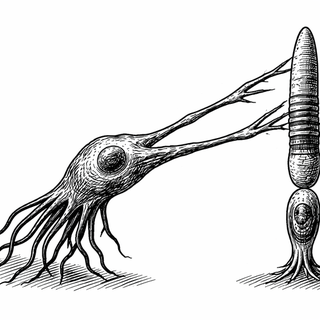
Most cells, when the floor drops out from under them, just... give up. Not the retinal pigment epithelium. When a genetic mutation rips open a gap between the RPE and the photoreceptors it's supposed to be feeding, these unassuming cells do something nobody predicted: they grow long, stretchy arms and reach across the void like a parent grabbing a toddler before it walks into traffic.
The Partnership That Keeps You Seeing
Here's a quick refresher on the most underappreciated relationship in your body. Your photoreceptors - the rods and cones that turn light into electrical signals - are constantly rebuilding themselves. Every day, they add fresh membrane at their base and shed old, worn-out material from their tips. The RPE, a single layer of cells sitting right behind your retina, acts as the cleanup crew, gobbling up those shed tips through a tightly choreographed process called phagocytosis (Lakkaraju et al., 2020).
Think of it like a conveyor belt at a sushi restaurant. The kitchen (photoreceptor base) keeps making new plates, old plates reach the end of the line, and somebody (the RPE) has to clear them away or the whole system jams. Scientists have always assumed this only works because the RPE sits right next to the photoreceptor tips - close enough to practically kiss them. Separate the two, and everything should fall apart.
Except it doesn't. Not right away, at least.
Enter the ADAM9 Knockout Mouse
ADAM9 is a metalloprotease - a protein that cuts other proteins - and when it's missing in humans, it causes cone-rod dystrophy, a progressive loss of vision (Parry et al., 2009). Mice lacking ADAM9 develop a dramatically expanded subretinal space, meaning the gap between their photoreceptors and RPE gets way bigger than it should be.
Clinicians have actually seen something similar in human diseases. In Best vitelliform macular dystrophy and age-related macular degeneration with subretinal drusenoid deposits, gunk accumulates between the photoreceptors and RPE, pushing them apart (Spaide et al., 2022). And here's the puzzle that's bugged researchers for years: patients with these conditions often maintain surprisingly good vision for a long time. The separation should be catastrophic, yet somehow the lights stay on.
The Stretch Armstrong Solution
Lewis and colleagues decided to look closely at what was actually happening in those ADAM9-knockout mouse retinas, and what they found was genuinely surprising (Lewis et al., 2026). Despite the expanded gap, photoreceptor light responses were completely normal. The rate of outer segment renewal? Also normal. The cells were working perfectly fine, thank you very much.
The secret? RPE cells were growing elongated pseudopods - finger-like projections - that stretched across the enlarged subretinal space to reach the photoreceptor tips and eat them, just like they normally would. These weren't subtle extensions. Using serial block-face scanning electron microscopy, the team captured RPE projections reaching dramatically farther than anyone thought possible.
As Akrit Sodhi put it in an accompanying commentary, this represents a fundamental "morphological plasticity" that challenges the proximity paradigm - the long-standing assumption that the RPE must be right next to photoreceptors to do its job (Sodhi, 2026).
What About the Immune Cells?
One prevailing theory had suggested that immune cells migrating into the subretinal space might be helping out, perhaps pitching in with the phagocytosis workload. The study found that wasn't the case. Those immune cells were just hanging around watching - spectators at a game they weren't playing in. The RPE was handling everything on its own, just with longer arms.
Why This Actually Matters
This isn't just a cool observation about mouse biology. It potentially explains something clinicians have wondered about for decades: why do patients with Best disease or early macular degeneration keep seeing so well despite visible separation between their photoreceptors and RPE?
The answer might be that human RPE cells can do the same stretching trick. If so, understanding what drives pseudopod formation - and what eventually causes it to fail - could open doors to therapies that keep this compensatory mechanism going longer. Delay the breakdown, delay the vision loss.
It also reframes how we think about the subretinal space itself. Rather than being a passive gap that spells doom when it expands, it's a space the RPE can actively navigate. These cells aren't just sitting there waiting for photoreceptor tips to come to them. When the situation demands it, they go get them.
Sometimes the most important discoveries aren't about finding new molecules or pathways. They're about watching cells do something nobody thought to look for - and realizing the eye had a Plan B all along.
References:
-
Lewis TR, Castillo CM, Phan S, et al. Adam9-deficient retinal pigment epithelium pseudopods maintain photoreceptor outer segment renewal despite subretinal space expansion. J Clin Invest. 2026;136(7):e196705. doi: 10.1172/JCI196705 | PubMed
-
Sodhi A. Retinal pigment epithelium pseudopods reach across the divide to maintain photoreceptor performance and save vision. J Clin Invest. 2026;136(7):e205127. doi: 10.1172/JCI205127
-
Parry DA, Toomes C, Senuber L, et al. Loss of the metalloprotease ADAM9 leads to cone-rod dystrophy in humans and retinal degeneration in mice. Am J Hum Genet. 2009;84(5):683-691. doi: 10.1016/j.ajhg.2009.04.005 | PMCID: PMC2681008
-
Lakkaraju A, Umapathy A, Tan LX, et al. The cell biology of the retinal pigment epithelium. Prog Retin Eye Res. 2020;78:100846. doi: 10.3389/fimmu.2020.604205
-
Spaide RF, Ooto S, Curcio CA. Subretinal drusenoid deposits AKA pseudodrusen. Surv Ophthalmol. 2018;63(6):782-815. doi: 10.1097/iae.0000000000003414 | PMCID: PMC9262011
Disclaimer: The image accompanying this article is for illustrative purposes only and does not depict actual experimental results, data, or biological mechanisms.