
Every decade past your twenties, your spinal cord quietly reshuffles its wiring - losing a bit of structure here, rewiring a connection there - and it does so in near-perfect lockstep with your brain, like two colleagues who never speak yet somehow always wear matching outfits.
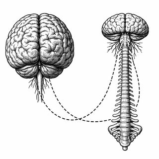
We tend to think of the spinal cord as a simple cable - the biological equivalent of a USB lead connecting your brain to everything south of your neck. Touch a hot stove, signal goes up; decide to move your hand, signal goes down. Terribly efficient, rather boring. Except it isn't boring at all. A new study published in Nature Communications by Landelle and colleagues has just demonstrated that the spinal cord has its own sophisticated structural and functional architecture, one that ages in patterns remarkably similar to the brain itself (Landelle et al., 2026).
The Cord Gets Its Close-Up
Here's the problem with studying the spinal cord: it's roughly the width of your little finger, it's surrounded by bone, and it moves every time you breathe. Imaging it with MRI is a bit like trying to photograph a hummingbird inside a washing machine. Most neuroscience studies have therefore politely ignored it and focused on the brain, which at least has the decency to sit still inside a rigid skull.
Landelle's team tackled this head-on (spine-on?) by assembling a multimodal dataset combining both spinal cord and brain imaging across a range of adults. They mapped the cord's microstructure using diffusion tensor imaging and captured its functional connectivity through resting-state fMRI - essentially eavesdropping on spontaneous neural chatter while participants lay quietly in the scanner, presumably wondering if they'd remembered to lock the front door.
What Ageing Actually Looks Like Down There
The findings paint a rather vivid picture of neural ageing. As the years tick by, the spinal cord shows microstructural decline - think of it as the insulation on the wiring gradually thinning. Functional connectivity shifts, and local spontaneous activity increases. The somatosensory pathway, which carries touch and body-position signals, took the biggest hit. This is the neural highway responsible for knowing where your feet are without looking at them, a skill you perhaps take for granted until, well, you can't.
Previous work had already established that individual spinal cord segments have their own organized resting-state networks, with motor regions in the front and sensory regions in the back showing distinct connectivity patterns (Kowalczyk et al., 2024). And earlier research from some of the same team had produced the first reliable functional parcellation of the cervical spinal cord, demonstrating that spinal "functional levels" could be mapped in individual people (Kinany, Landelle et al., 2024). But nobody had tracked how all of this changes as we age - or whether those changes mirror what happens upstairs.
Your Brain and Spine: Same Memo, Different Office
The really striking bit is the convergence. When the team extended their analysis to include brain imaging, they found the same ageing signatures in both structures: gray matter loss, decreased functional segregation (brain networks becoming less distinct from one another, like radio stations drifting onto the same frequency), and increased spontaneous activity. It's a CNS-wide phenomenon - your brain and spinal cord aren't just connected, they're ageing on the same schedule with the same playbook.
This matters because it suggests that what we observe in brain ageing studies isn't merely a cortical affair. The entire central nervous system appears to follow shared biological trajectories. Research into brain network segregation during ageing has shown that when functional networks lose their distinctiveness, cognitive decline often follows (Chan et al., 2020). Now we know the cord is playing the same tune.
Why Should You Care (Besides General Existential Dread)?
If spinal cord changes track with brain changes, the cord could serve as an accessible imaging marker for broader neural ageing. Think of it as a canary in a neurological coal mine. Spinal imaging might one day help clinicians detect early sensorimotor decline before someone notices their balance is off or their grip is weakening.
For now, this study provides the first systems-level map of how the brain and spinal cord age together - a foundation that future research on conditions like multiple sclerosis, spinal cord injury, and age-related sensorimotor dysfunction can build upon. It's a reminder that the most underappreciated cable in your body has been running a rather complex operation this entire time, and it deserves the same scientific attention we lavish on the brain.
Your spine, it turns out, has been keeping pace with your brain all along. It's just too polite to mention it.
References
-
Landelle, C., Kinany, N., St-Onge, S., Lungu, O., Van De Ville, D., Misic, B., Marchand-Pauvert, V., De Leener, B., & Doyon, J. (2026). Spinal cord structural and functional architecture and its shared organization with the brain across the adult lifespan. Nature Communications. https://doi.org/10.1038/s41467-026-71963-2
-
Kowalczyk, O. S., Payne, S., Engel, M., Tinnermann, A., Nahman-Averbuch, H., & Brooks, J. C. W. (2024). Spinal fMRI demonstrates segmental organisation of functionally connected networks in the cervical spinal cord: A test-retest reliability study. Human Brain Mapping, 45(2), e26600. https://doi.org/10.1002/hbm.26600
-
Kinany, N., Landelle, C., Kinany, N., et al. (2024). In vivo parcellation of the human spinal cord functional architecture. Imaging Neuroscience, 2, 1-17. https://doi.org/10.1162/imag_a_00059
-
Chan, M. Y., Park, D. C., Savalia, N. K., Petersen, S. E., & Wig, G. S. (2020). Decreased segregation of brain systems across the healthy adult lifespan. Proceedings of the National Academy of Sciences. https://doi.org/10.1007/s12264-020-00455-2
Disclaimer: The image accompanying this article is for illustrative purposes only and does not depict actual experimental results, data, or biological mechanisms.