
Bad timing rewires vision.
That is the basic scam behind amblyopia. If one eye delivers lousy input during early development, the brain starts treating that eye like the intern who keeps crashing the shared spreadsheet. It stops trusting it. The result is not just an eye problem - it is a wiring problem in the visual brain. The new mouse study behind this post asks a gloriously weird question: what if you could reboot that wiring by briefly silencing the retina itself?
The old product failed
Classic amblyopia treatment usually means glasses, patching the stronger eye, or blurring it with atropine so the weaker eye has to do some work. That can help, especially in kids, but it is not exactly a frictionless user experience. Children often hate patching with the passion of a thousand suns, and treatment gets less reliable as the brain gets older. Deprivation amblyopia, caused by early blockage of vision such as cataract, is especially stubborn [2-4].
The big idea in the new paper is that maybe recovery is not only about forcing the weak eye to perform. Maybe it is about changing the brain state that decides whether visual connections can strengthen at all. In startup language, the team is not just tweaking the app UI. They are trying to reset the recommendation engine.
What the mice showed
The study looked at mice that had undergone long-term monocular deprivation during the critical period, a standard model of amblyopia. The researchers temporarily inactivated one retina with tetrodotoxin, or TTX, a toxin that blocks action potentials. Very chill molecule. Extremely not DIY.
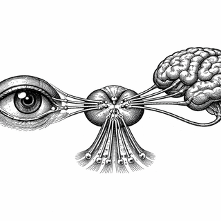
Here is the key result. When one eye was silenced, neurons in the dorsal lateral geniculate nucleus, or dLGN, started firing in high-frequency bursts. The dLGN is the thalamic relay between the eye and visual cortex, so think of it as the overworked transit hub through which visual information commutes before it hits cortex. Those bursts depended on low-threshold T-type calcium channels, especially CaV3.1. When the researchers genetically removed CaV3.1 in the dLGN, the therapeutic effect disappeared [1].
That matters because it points to a mechanism, not just a cool outcome. The recovery seemed to require a specific firing mode in the thalamus. Burst firing may act like a giant "plasticity is open" sign for cortex, helping weakened eye inputs strengthen again.
And then came the plot twist. Prior work had emphasized silencing the fellow eye, the stronger one. But in this study, silencing the amblyopic eye alone also drove recovery. That suggests the burst pattern itself may be the important ingredient, not simply depriving the better eye of competition [1]. The thalamus, that deeply unglamorous middle manager, may have been quietly running the turnaround plan the whole time.
Why this is more than a rodent party trick
This paper lands in a bigger shift in amblyopia science. The old dogma said the adult visual system was basically done shipping updates after childhood. Newer work has been chipping away at that for years. Reviews, trials, and clinical guidance now argue that treatment can still work later than once believed, and that binocular approaches deserve serious attention alongside patching [2-4].
At the same time, basic neuroscience has kept showing that ocular dominance plasticity is not dead in adulthood - it is just harder to coax out, like a software team after its fourth reorg. Work in visual cortex has identified circuit-level gates on this plasticity [5]. The new Cell Reports paper adds the thalamus to the VIP list. Not just cortex. Not just eye. The relay station in between may be setting the tone.
The clinical dream, with several asterisks
If this mechanism holds up, the long-term implication is obvious: amblyopia treatment might someday aim to trigger the right plasticity state rather than merely punish the stronger eye. That could matter for older patients, for severe deprivation amblyopia, and for people who do not respond well to standard patching.
But let’s keep both shoes on the ground. This is a mouse study. It measured cortical responses, not a full human visual comeback story. TTX retinal inactivation is also not a ready-made clinic procedure. The challenge now is finding safer ways to reproduce the beneficial signal without causing harm.
Still, the concept is deliciously provocative. Amblyopia may not be a permanently failed product after all. It may be a system stuck in the wrong mode, waiting for the right reboot command. And if the thalamus really is the switchboard operator here, neuroscience once again delivers the humbling news that the weird middle layer nobody was bragging about might be the one keeping the whole company alive.
References
- Echavarri-Leet M, Chauhan T, Cramer TLM, Fong MF, Bear MF. Temporary retinal inactivation reverses effects of long-term monocular deprivation in visual cortex by induction of burst mode firing in the thalamus. Cell Reports. 2025;45:116566. DOI: 10.1016/j.celrep.2025.116566. PubMed: 41240337.
- Proudlock FA, Gottlob I. Are we any closer to optimising amblyopia treatment? Clinical and Translational Medicine. 2024;14(12):e70080. DOI: 10.1002/ctm2.70080. PMCID: PMC11608889.
- Proudlock FA, Hisaund M, Maconachie G, et al. Extended optical treatment versus early patching with an intensive patching regimen in children with amblyopia in Europe (EuPatch): a multicentre, randomised controlled trial. The Lancet. 2024;403(10438):1766-1778. DOI: 10.1016/S0140-6736(23)02893-3. PubMed: 38704172.
- Cruz OA, Repka MX, Hercinovic A, et al. Amblyopia preferred practice pattern. Ophthalmology. 2023;130(3):P136-P178. DOI: 10.1016/j.ophtha.2022.11.003. PMCID: PMC10260971.
- Severin D, Hong SZ, Roh SE, et al. All-or-none disconnection of pyramidal inputs onto parvalbumin-positive interneurons gates ocular dominance plasticity. Proceedings of the National Academy of Sciences of the United States of America. 2021;118(37):e2105388118. DOI: 10.1073/pnas.2105388118. PubMed: 34508001.
Disclaimer: The image accompanying this article is for illustrative purposes only and does not depict actual experimental results, data, or biological mechanisms.