
That matters because multiple sclerosis is usually told as a story about the immune system picking a fight with the brain; this paper suggests the brain may also be quietly texting the immune system from headquarters, and in MS that group chat starts malfunctioning. If Plato had written about autoimmune disease, he probably would have blamed the shadows and then gone looking for the hypothalamus.
In a new Cell Reports study, Tiziana Vigo and colleagues followed one of those odd backchannels: hypothalamic AgRP neurons, noradrenergic sympathetic fibers, the bone marrow, and the thymus. In mice with experimental autoimmune encephalomyelitis, or EAE, a standard model of MS, they found that this brain-to-immune relay helps control blood-cell production in bone marrow and the balance of regulatory T cells in the thymus through beta-3 adrenergic receptors. Then came the unpleasant plot twist: in EAE, that relay was impaired, and in people with MS, higher blood AgRP levels tracked with worse disease severity and MRI signs of neuroinflammation (1).
The Brain Is Not Just Brooding
AgRP neurons are usually famous for hunger. They are the brain cells most likely to insist that shredded cheese counts as a meal. But the hypothalamus does more than manage appetite; it also helps run stress responses and autonomic output to the body.
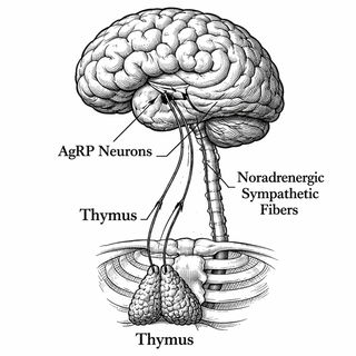
The broader idea is not brand new: the nervous and immune systems constantly negotiate through sympathetic nerves and molecules like norepinephrine (2, 3). What is new is the route. The authors place AgRP neurons upstream of noradrenergic signaling in two immune factories: bone marrow, where blood cells are born, and the thymus, where T cells mature and ideally learn some restraint.
Bone Marrow, Thymus, and Other Places You Forget Exist
Bone marrow sounds like medieval soup, but it is really the body’s blood-cell workshop. Stem and progenitor cells develop inside organized niches shaped by stromal cells, vessels, and nerves (4). The thymus, meanwhile, is the finishing school for T cells, including regulatory T cells, or Tregs, whose main job is stopping the immune system from behaving like a comment section (5).
Vigo and colleagues found that noradrenergic signaling pushed the bone marrow toward myeloid hematopoiesis and influenced the fraction of Tregs in the thymus through beta-3 adrenergic receptors. When AgRP neurons were dysfunctional in EAE, that control weakened. So MS-like inflammation may not just damage brain tissue directly; it may also distort how the brain instructs immune organs to stock the shelves. The disease might be changing the command center while also changing the troops.
Why This Is Interesting Beyond Mouse Drama
MS research has been moving toward a more connected picture of disease: immune cells, glia, neurons, brain borders, peripheral tissues, and repair mechanisms all seem to be in one sprawling argument rather than neat separate chapters (6, 7). In February 2025, NIH researchers also reported an animal “4D brain map” that may help spot lesion-prone regions before visible MS-like damage appears, which tells you the field is leaning toward earlier, more network-aware ways of understanding disease (8).
This paper fits that shift. It says, in effect, maybe the brain is not merely the victim of immune chaos; maybe it also helps tune the immune landscape in advance. If that holds up, therapies might eventually target not only immune cells but also the neural circuits and adrenergic signals that shape how those cells are produced and educated.
The Catch, Because There Is Always a Catch
Before anybody starts naming a biotech startup after AgRP, the cautions matter. Most of the mechanistic work here was done in mice with EAE, which is useful but not identical to human MS. The human side is correlational: higher serum AgRP tracked with worse disease and MRI measures, but correlation is a flirtation, not a marriage certificate.
There is also the usual receptor-targeting headache. Beta-adrenergic pathways show up in many tissues, and the sympathetic nervous system is not exactly a boutique signal confined to one tidy organ. If you tug on this circuitry therapeutically, you would want precision.
Still, the challenge this work addresses is real: MS is not just about stopping immune attacks, but understanding why the immune system gets the wrong instructions in the first place, and why repair remains so incomplete. A brain-controlled immune relay linking appetite-related hypothalamic neurons to bone marrow and thymus was not on many people’s bingo cards; now it probably should be.
References
- Vigo T, Mariani MC, Bason C, et al. Noradrenergic control of bone marrow and thymus by AgRP neurons is impaired in experimental multiple sclerosis. Cell Reports. 2025;44(11):116556. DOI: https://doi.org/10.1016/j.celrep.2025.116556
- Cao Y, Chen H, Yang J. Neuroanatomy of lymphoid organs: Lessons learned from whole-tissue imaging studies. European Journal of Immunology. 2023;53(10):e2250136. DOI: https://doi.org/10.1002/eji.202250136
- Gentile A, Musella A, Fresegna D, et al. The distinct roles of monoamines in multiple sclerosis: A bridge between the immune and nervous systems? Brain, Behavior, and Immunity. 2021;94:254-264. DOI: https://doi.org/10.1016/j.bbi.2021.02.030
- Lucas D. Structural organization of the bone marrow and its role in hematopoiesis. Current Opinion in Hematology. 2021;28(1):36-42. DOI: https://doi.org/10.1097/MOH.0000000000000621. PMCID: https://pmc.ncbi.nlm.nih.gov/articles/PMC7769132/
- Marx A, Yamada Y, Simon-Keller K, et al. Thymus and autoimmunity. Seminars in Immunopathology. 2021;43(1):45-64. DOI: https://doi.org/10.1007/s00281-021-00842-3
- Attfield KE, Jensen LT, Kaufmann M, Friese MA, Fugger L. The immunology of multiple sclerosis. Nature Reviews Immunology. 2022;22:734-750. DOI: https://doi.org/10.1038/s41577-022-00718-z
- Woo MS, Engler JB, Friese MA. The neuropathobiology of multiple sclerosis. Nature Reviews Neuroscience. 2024;25:493-513. DOI: https://doi.org/10.1038/s41583-024-00823-z
- Lin J-P, et al. 4D marmoset brain map reveals MRI and molecular signatures for onset of multiple sclerosis-like lesions. Science. 2025. DOI: https://doi.org/10.1126/science.adp6325
Disclaimer: The image accompanying this article is for illustrative purposes only and does not depict actual experimental results, data, or biological mechanisms.