That, more or less, is the game in this new paper. And it is a surprisingly ambitious one.
Neuroscience has a long-standing habit of learning things in mice and then looking terribly hopeful about humans afterward. Fair enough - mice are small, genetically tractable, and less likely to complain to the ethics board in complete sentences. But the awkward truth has always been this: a mouse brain is not just a tiny human brain with whiskers. If a treatment works in mice but the mouse model only vaguely resembles the human disease, you may as well be testing umbrella design on a haddock.
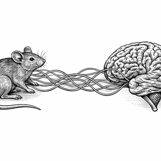
This study, by Jaroszynski and colleagues, tries to fix that translation problem with something rather elegant. They used spatial transcriptomics - measurements of which genes are active in different brain regions - and trained a variational autoencoder, a machine-learning model that compresses complicated data into a more manageable "latent space." Which sounds suspiciously like something a tech founder would say before asking for £40 million, but here it is genuinely useful. The model learned a shared gene-expression landscape for mouse and human brains, using orthologous genes - roughly, genes in different species descended from the same ancestral gene.
A bilingual dictionary for brains
The basic idea is this: instead of matching mouse and human brains only by gross anatomy - this lobe, that nucleus, everyone nod politely - the authors matched them by deeper molecular patterns. Brain regions from both species that shared similar latent gene-expression signatures ended up close together in this common space.
And reassuringly, the model did not immediately sprint into nonsense. Known anatomical counterparts tended to align better in this latent space, and broad organisational principles were preserved across species. In other words, the system seemed to recover real biology rather than inventing modern art.
That matters because cross-species neuroscience often lives in a foggy middle ground. We know mice are useful. We know they are imperfect. We also know that "imperfect" is doing heavy lifting there. A formal quantitative framework for asking which mouse brain regions best match which human regions is far more helpful than the traditional method, which is sometimes just "well, the hippocampus looks familiar enough."
Related work has been pushing in this direction. Large cross-species cell atlases and spatial transcriptomic maps have shown that gene-expression patterns can reveal evolutionary continuity and divergence with much finer resolution than old-school anatomy alone (Hodge et al., 2023, Nature, DOI: 10.1038/s41586-023-05812-9; Yao et al., 2023, Nature, DOI: 10.1038/s41586-023-06812-5). Which is encouraging, because the brain has always been rather rude about looking similar while doing subtly different things.
Alzheimer’s, Parkinson’s, and the annual mouse-model beauty contest
The clever bit comes next. The authors did not stop at anatomy. They asked whether disease-related brain changes in mouse models could be translated into predicted human patterns - specifically for Alzheimer’s disease and Parkinson’s disease.
And the answer was: to a meaningful extent, yes.
Their framework could take alterations seen in mouse disease models and generate patterns that matched known human brain changes. For Alzheimer’s in particular, they could compare multiple mouse models and different timepoints, then ask which ones best resembled patient data. This is quietly radical. Instead of treating "mouse model of Alzheimer’s" as a single category, like calling every pasta shape "basically spaghetti," the method helps sort which models fit which stage or aspect of the disease.
That could matter a great deal in drug development, where a depressingly large number of promising preclinical results vanish on contact with human trials. Reviews over the past few years have repeatedly pointed to poor model validity as a major bottleneck in neurodegenerative research (Scearce-Levie et al., 2020, Neuron, DOI: 10.1016/j.neuron.2020.11.034; De Strooper and Karran, 2023, Cell, DOI: 10.1016/j.cell.2023.02.031). If you can choose a model because it molecularly maps onto the relevant human disease stage, you are no longer throwing darts in the dark. You are still throwing darts, because biology enjoys humiliation as a hobby, but at least the lights are on.
Why this is more than a neat computational trick
This paper lands in a bigger moment for neuroscience. Spatial transcriptomics has exploded because it lets researchers ask not just what genes are active, but where in tissue they are active. That "where" is everything in the brain, where location is destiny and neighbouring cells can have utterly different jobs, personalities, and probable opinions about the prefrontal cortex.
A translational framework built on these maps could help with:
- picking better animal models before expensive studies begin
- identifying which disease stage a model actually resembles
- interpreting why some therapies fail when moved into people
- narrowing the gap between basic neuroscience and clinical trials
Of course, there are caveats. Mouse and human brains differ in lifespan, scale, connectivity, and behaviour in ways no latent space can magic away. Gene-expression similarity is powerful, but it is not the whole story. Brains are not just bags of transcripts with opinions. They are shaped by development, experience, cell interactions, and all the other untidy details that make biology so charmingly uncooperative.
Still, this is one of those papers that changes the question. Not "is the mouse useful?" but "useful for which human problem, exactly?" That is a much better question. It may also save everyone a great deal of time, money, and dignified disappointment.
References
Jaroszynski C, Amer M, Beauchamp A, Lerch JP, Sotiropoulos SN, Mars RB. Translating brain anatomy and disease from mouse to human in latent gene expression space. EBioMedicine. 2026;106259. doi:10.1016/j.ebiom.2026.106259
Hodge RD, et al. Conserved cell types with divergent features in human versus mouse cortex. Nature. 2023. doi:10.1038/s41586-023-05812-9
Yao Z, et al. A high-resolution transcriptomic and spatial atlas of cell types in the whole mouse brain. Nature. 2023. doi:10.1038/s41586-023-06812-5
Scearce-Levie K, et al. Improving preclinical models of neurodegenerative disease. Neuron. 2020. doi:10.1016/j.neuron.2020.11.034
De Strooper B, Karran E. The cellular phase of Alzheimer’s disease. Cell. 2023. doi:10.1016/j.cell.2023.02.031
Disclaimer: The image accompanying this article is for illustrative purposes only and does not depict actual experimental results, data, or biological mechanisms.