
You're using this right now. Every letter on this screen is barging into your eyes at once, and yet your visual system is not collapsing into the neurological equivalent of a pub quiz team shouting four answers at the same time. That is partly because neurons use a trick called normalization - a way of keeping responses from blowing up when the world gets visually crowded. In a new monkey study, researchers asked a sneaky question: when multiple motion signals land in different parts of a neuron's turf, does the brain average them evenly, or does location quietly rig the vote?
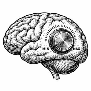
Normalization is one of those ideas in neuroscience that sounds dry until you realize it is basically constant damage control. A neuron in visual cortex does not just add up every input like a cashier with a broken calculator. It scales its response based on what else is happening nearby, which helps preserve selectivity instead of letting a pile of mediocre signals impersonate one great one (Goris et al., 2024; PMC11444047).
The new paper focuses on area MT, a motion-sensitive patch of monkey visual cortex. MT neurons care a lot about where and how things move. They also have fairly large receptive fields, meaning each neuron listens to a decent chunk of visual space rather than one tiny pixel of existence. That matters because real scenes are cluttered.
Not all parts of a receptive field are created equal
Cherian and Maunsell recorded from 90 MT neurons in macaque monkeys and flashed moving Gabor patches at different positions inside each neuron's receptive field while the animals did a separate change-detection task. One stimulus sat near the receptive-field center, while another could appear farther out on the flank. The question was simple: if two stimuli show up, how much does each one count?
Classic normalization models did not handle these arbitrary positions very well. The better fit was an "intensity-weighted" model, where intensity means stimulus contrast multiplied by a location-specific receptive-field weight. In plain English, a stimulus counts more if it is strong and if it lands where the neuron actually cares.
That tweak also explained why contrast sensitivity changed with location. Stimuli closer to the receptive-field center were not just better at driving the neuron. They also changed the normalization pool more strongly. Even the neuron's so-called spontaneous activity looked less like random static and more like an extra source of excitatory drive that joins the same accounting system. The background noise, apparently, was on the committee.
Why this matters outside the monkey lab
If this result holds up and generalizes, it sharpens an old idea into something more realistic. The brain may not normalize visual inputs by contrast alone. It may normalize by effective intensity, which depends on where the signal lands in the neuron's receptive field and how much that neuron is set up to care about that location. Natural vision is mostly clutter management, and this gives a cleaner rule for how the system keeps order.
Recent work lines up with that broader picture. A 2021 PNAS study argued that divisive normalization can unify response patterns across the human visual hierarchy, not just in one tiny corner of cortex (Aqil et al., 2021; PMC8609633). A 2024 Nature Communications paper showed that stable contrast processing in dynamic natural scenes can emerge from normalization-like circuitry, this time in flies (Gur et al., 2024).
There is also a practical reason to care. Area MT and related motion systems matter for navigation, balance, and interpreting a moving world. When motion processing goes wrong, people can get debilitating symptoms, including motion blindness, or akinetopsia (Sallam et al., 2024). Better models of how visual neurons stabilize and rank competing motion signals could eventually help researchers think more clearly about motion-perception disorders and brain-inspired computer vision that does not panic when the background gets busy.
The nice part: it makes the theory less magical
One quiet win in this paper is that it makes normalization feel less like a mystery constant hiding in an equation and more like a physical story about weighted inputs. The brain is still a wet electric gossip network that refuses to explain itself in a straight line. But this study gives a cleaner rule for one of its staple tricks: when MT neurons combine motion signals, they seem to average by effective intensity, not by raw contrast alone. In a world full of visual clutter, that is exactly the sort of fussy bookkeeping you would want.
References
Cherian C, Maunsell JHR. Neuronal normalization in monkey MT is an intensity-weighted average. Proc Natl Acad Sci U S A. 2025;122(45):e2522104122. doi:10.1073/pnas.2522104122. PubMed:41196346.
Goris RLT, Coen-Cagli R, Miller KD, Priebe NJ, Lengyel M. Response sub-additivity and variability quenching in visual cortex. Nat Rev Neurosci. 2024;25(4):237-252. doi:10.1038/s41583-024-00795-0. PMCID:PMC11444047.
Aqil M, Knapen T, Dumoulin SO. Divisive normalization unifies disparate response signatures throughout the human visual hierarchy. Proc Natl Acad Sci U S A. 2021;118(46):e2108713118. doi:10.1073/pnas.2108713118. PMCID:PMC8609633.
Gur B, He Q, Munch TA, et al. Neural pathways and computations that achieve stable contrast processing tuned to natural scenes. Nat Commun. 2024;15(1):8580. doi:10.1038/s41467-024-52724-5.
Sallam A, et al. Akinetopsia: a systematic review on visual motion blindness. Front Neurol. 2024. doi:10.3389/fneur.2024.1510807.
Disclaimer: The image accompanying this article is for illustrative purposes only and does not depict actual experimental results, data, or biological mechanisms.