Microns are rude. A single vagal sensory neuron is so tiny you could fit a small army of them across the width of a human hair, yet one odd little subgroup may help decide whether a signal from your gut gets treated like a boring household memo or a VIP backstage pass to the brain's reward system.
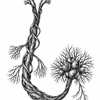
The paper by Welch and colleagues looks at the vagus nerve, the long wandering bundle that carries updates from your organs to your brain. A lot of that traffic comes from the gut. Full stomach? Weird nutrient mix? Hormones released during a meal? The vagus is on it.
Scientists already had a strong clue that the right vagus nerve plays a special role in reward-related signaling from the gut. Earlier work suggested that stimulating gut-linked vagal pathways can drive dopamine release and reinforce appetitive behavior, which is neuroscience-speak for "the body can make food feel extra worth pursuing" Han et al., 2018. More recent reviews have pushed the field away from the old idea that vagal sensory neurons are just passive fullness detectors Décarie-Spain et al., 2024, Mendez-Hernandez et al., 2025.
Right Side, Main Character Energy
So what did this new study find? Using single-nucleus RNA sequencing, RNA labeling in tissue, and calcium imaging, the researchers identified a specific set of vagal sensory neurons that co-express Chrna3 and Cckar. Those cells showed up more on the right side of the nodose ganglion, which is the sensory ganglion that houses many vagal neuron cell bodies. In mice and rats, these right-biased neurons also expressed genes such as Glp1r and Sctr, which makes them look a lot like gut-focused chemical sensors rather than generic all-purpose messengers Welch et al., 2025.
That matters because Cckar encodes the receptor for cholecystokinin, or CCK, a gut hormone released during meals. CCK has long been famous for helping signal satiation, basically the body's polite-but-firm "we can stop eating now" notice. But in the right neural context, meal-related gut signals may do more than tap the brakes. They may also help tag food-related experiences as rewarding and worth repeating.
Why This Is More Than Left Versus Right Trivia
At first glance, "some neurons are more common on the right than the left" sounds like trivia. But this can explain why whole circuits behave differently. If the right vagus contains more of a neuron type tuned to digestive hormones and nutrient-related chemistry, that gives researchers a concrete cellular suspect for asymmetric gut-brain reward signaling.
Think of it this way: if two kids have walkie-talkies but only one of them has the direct line to the snack table, you should not act surprised when that kid suddenly becomes very influential.
This also helps with a broader puzzle in neuroscience called interoception, which is the brain's running readout of the body's internal state. Hunger, nausea, fullness, calm, that ominous "I ate too fast and now regret my life choices" feeling - those all depend on internal sensing. The vagus nerve is one of the major routes for that information, and this paper sharpens the map by showing that the route is not perfectly symmetrical Wikipedia: Vagus nerve, Wikipedia: Interoception.
The Big Promise, With the Usual Fine Print
If these findings hold up and expand, they could matter for conditions where reward, appetite, and gut signaling get tangled together - obesity, binge eating, disordered eating, even addiction-related processes. A 2026 study in Science Advances adds weight to that possibility by showing that gut-brain vagal signaling helps govern mesolimbic dopamine dynamics and reward events more broadly Onimus et al., 2026. That does not mean your vagus nerve is secretly a tiny nightclub promoter for every craving you have ever had. It does mean the old brain-only story about reward looks less complete.
The catch is that this Cell Reports study is still an early mechanistic step. It identified a compelling right-biased cell population and showed functional asymmetry in response to CCK-related signaling, but it did not fully prove that these exact neurons are sufficient to drive reward behavior on their own.
Still, this is the good kind of complication. It replaces a vague idea with a testable one: a specific molecularly defined neuron group on the right side may carry a special piece of the gut-to-reward conversation.
And honestly, the brain deserves some humility here. For years it acted like the sole executive in charge. Meanwhile, the gut kept sending notes from downstairs, and now we are discovering that one side of the body's wandering nerve may have been slipping dopamine-relevant messages into the inbox all along.
References
- Welch HF, et al. Cell Reports (2025). DOI: https://doi.org/10.1016/j.celrep.2025.116507 | PubMed: https://pubmed.ncbi.nlm.nih.gov/41196683/
- Décarie-Spain L, et al. Semin Cell Dev Biol (2024). DOI: https://doi.org/10.1016/j.semcdb.2023.02.004 | PMCID: https://pmc.ncbi.nlm.nih.gov/articles/PMC10427741/
- Mendez-Hernandez R, et al. Compr Physiol (2025). DOI: https://doi.org/10.1002/cph4.70010 | PMCID: https://pmc.ncbi.nlm.nih.gov/articles/PMC12090708/
- Onimus O, et al. Science Advances (2026). DOI: https://doi.org/10.1126/sciadv.adz0828 | PMCID: https://pmc.ncbi.nlm.nih.gov/articles/PMC12857734/
- Han W, et al. Cell (2018). DOI: https://doi.org/10.1016/j.cell.2018.08.049 | PubMed: https://pubmed.ncbi.nlm.nih.gov/30245012/
Disclaimer: The image accompanying this article is for illustrative purposes only and does not depict actual experimental results, data, or biological mechanisms.