
The surprise in Budoff and Poleg-Polsky's data was not that mouse retinal ganglion cells have favorite neighborhoods. People already suspected some of that. The real eyebrow-launcher was that some cells with the same molecular identity seemed to change their gene expression depending on where they lived. Same cell type, different postal code, different vibe. Your retina, it seems, is less like a tidy spreadsheet and more like a city where identical coffee shops somehow become completely different after crossing one street.
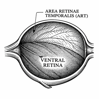
That matters because retinal ganglion cells, or RGCs, are the retina's output neurons. They are the last stop before visual information leaves the eye and heads for the brain. If the retina is a layered little computing device, RGCs are the cables carrying its conclusions. They are not all doing the same job, either. Some care about motion, some about contrast, some about light level, and some are basically the eye's internal clock support staff. Very overbooked, slightly chaotic, deeply neuronal.
A map, but make it weird
In this 2025 PNAS paper, the authors used spatial transcriptomics, en-face cryosectioning, and machine learning to build a retina-wide map of mouse RGC types. Earlier work had identified many RGC types from single-cell RNA sequencing, but that approach removes cells from the tissue. Great for reading molecular name tags, bad for knowing where everybody was standing before the party got broken up.
This study put the cells back on the floor plan.
About two-thirds of RGC types were spread fairly evenly across the retina. Nice. Polite. Euclidean. But the rest showed clear spatial biases, especially toward the ventral retina, which samples the sky, and toward a dorso-temporal region called the area retinae temporalis, or ART. Think of the ART as the mouse retina's premium seating section. Not a full primate-style macula, not the velvet rope at Studio 54, but definitely not random.
Then came the plot twist. Some RGC types did not just vary in number from place to place. Their gene expression also shifted across regions, including along the dorsal-ventral axis and inside versus outside the ART. That means location is not merely population math. It may tune what a cell is, or at least what molecular tools it keeps in the toolbox. Geometry and identity are not separable here. The retina is doing topology before breakfast.
Why should you care if a mouse sees the sky differently?
Because vision is not uniform, and neither are the problems that break it.
RGCs are the cells that die in glaucoma and other optic nerve diseases. If some RGC types cluster in certain regions, or if the same type behaves differently depending on where it sits, then vulnerability to disease might also follow a map rather than a simple list. That idea already fits the direction of the field: a 2025 Neuron study using flatmount MERFISH found that many mouse RGC types are spatially biased and that some cells near blood vessels appear more resilient in glaucoma-like conditions. In other words, retinal real estate might matter more than neuroscientists would prefer, which is rude but informative.
There is also a basic science payoff. The retina often gets described as a camera sensor, but that undersells it badly. It is more like a compressed sensing algorithm with opinions. Different parts of visual space matter differently to an animal. For a mouse, the upper visual field can mean predators. So if certain RGC types are enriched where sky information lands, that is not decorative biology. It is ecological prioritization with cellular paperwork.
Not just a mouse version of the human eye
One especially useful result is what the paper did not find. The authors looked for similarities between the mouse ART and the primate macula, the high-acuity center of primate vision, and found only limited correlation in gene-expression profiles. So the mouse ART is not a tiny discount macula with a fake mustache.
That is important because mouse vision research often tempts us into easy analogies. Mice are invaluable, but evolution is not a photocopier. Sometimes it uses the same trick. Sometimes it solves the same problem with a completely different circuit and hopes nobody notices. This paper politely notices.
The bigger challenge hiding underneath
The hard problem here is that cell type is not a single label. It is a bundle of function, shape, connectivity, gene expression, and now, more clearly than before, spatial context. Neuroscience loves categories because categories make us feel in control, the way organizing cables makes you feel like a person who definitely has their life together. Then biology walks in holding a box of unlabeled adapters.
Recent atlases and classification studies have been pushing toward this richer view of retinal organization, from unified RGC type schemes to whole-retina MERFISH maps. Budoff and Poleg-Polsky add something sharp to that picture: even once you think you know the cast of characters, you still need the seating chart.
If the result holds up and expands across development, disease, and species, it could help researchers design more precise experiments, interpret retinal degeneration more intelligently, and maybe one day target therapies to the cells and regions that need them most. That is the practical promise. The conceptual one is even nicer: the retina is not just a list of parts. It is a patterned surface where location helps write the rules.
Which, honestly, is exactly the kind of move the brain loves - take a hard problem, turn it into a map, then hide extra complexity in the map anyway. Classic.
References
- Budoff SA, Poleg-Polsky A. A complete spatial map of mouse retinal ganglion cells reveals density and gene expression specializations. Proc Natl Acad Sci U S A. 2025;122(52):e2515449122. DOI: 10.1073/pnas.2515449122. PubMed: 41452983
- Goetz J, Jessen ZF, Jacobi A, et al. Unified classification of mouse retinal ganglion cells using function, morphology, and gene expression. Cell Reports. 2022;40(5):111040. DOI: 10.1016/j.celrep.2022.111040
- Choi J, Li J, Ferdous S, et al. Spatial organization of the mouse retina at single cell resolution by MERFISH. Nature Communications. 2023;14:4929. DOI: 10.1038/s41467-023-40674-3
- Tian L, Chen F, Macosko EZ. The expanding vistas of spatial transcriptomics. Nature Biotechnology. 2023;41(6):773-782. DOI: 10.1038/s41587-022-01448-2. PMCID: PMC10091579
- Piwecka M, Rajewsky N, Rybak-Wolf A. Single-cell and spatial transcriptomics: deciphering brain complexity in health and disease. Nature Reviews Neurology. 2023;19(6):346-362. DOI: 10.1038/s41582-023-00809-y. PMCID: PMC10191412
- Nimkar K, Tsai NY, Zhao M, et al. Molecular and spatial analysis of ganglion cells on retinal flatmounts identifies perivascular neurons resilient to glaucoma. Neuron. 2025;113(20):3390-3407.e8. DOI: 10.1016/j.neuron.2025.07.025. PubMed: 40840447
Disclaimer: The image accompanying this article is for illustrative purposes only and does not depict actual experimental results, data, or biological mechanisms.