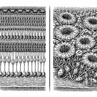
Under a microscope, the healthy retina looks almost absurdly tidy - thin bands of cells stacked like pastry layers, each one crisp, deliberate, and a little smug about its organizational skills. In the new eLife paper Afadin sorts different retinal neuron types into accurate cellular layers, that neat arrangement starts to fray. Knock out a protein called Afadin in developing mouse retina, and the whole place begins to look less like elegant architecture and more like a theater where everyone ignored their assigned seats.
That matters because your retina is not just wallpaper at the back of the eye. It is a living circuit board that starts processing visual information before the signal ever reaches the brain. The retina works partly because its cell types live in the right places and meet the right partners, not because the cells simply exist at all (Wikipedia: Retina).
Not Just Glue, More Like a Very Opinionated Stage Manager
Afadin is a cytosolic adaptor protein. In plain English, it helps connect cell-adhesion molecules at the cell surface to the internal actin skeleton. That sounds dry until you realize what it means: cells are not only sticking to things, they are translating those sticky handshakes into movement, shape, and final position. Afadin is part of the backstage crew that turns “you belong near me” into “please stand exactly here and do not freeload into the wrong layer.”
Lum and colleagues used conditional genetics in mice to remove Afadin during retinal development. The result was not a mass die-off of neurons. The cells still formed major classes such as retinal ganglion cells, bipolar cells, and amacrine cells. But many of them ended up in the wrong layers, with severe lamination defects, rosette-like structures, and neurons bridging spaces they normally should not cross (Lum et al., 2026). That is the plot twist: the retina can make the right cast but still botch the seating chart.
Why the Seating Chart Exists in the First Place
Retinal layering is not decorative. It is how the tissue keeps specific conversations from turning into a neurological group chat from hell. If those cell types drift, the geometry of who can talk to whom changes too.
Developmental neuroscientists have known for a while that retinal wiring depends on a mix of cell identity, migration, adhesion, and timing. Reviews over the last five years have emphasized that retinal neurons do not just pick partners chemically, they also rely on layer-specific positioning and adhesion systems to build circuits with any hope of precision (Malin and Desplan, 2021; Graham and Duan, 2020; D'Souza and Lang, 2020).
This new study sharpens that picture. It says Afadin is not merely helping cells stay attached in a vague, “biology likes cohesion” way. It appears to help different retinal neuron types sort into the proper laminae while preserving the basic identity of those neurons. That is a more specific job description, and biology loves a protein with a weirdly niche side hustle.
The Weirdly Reassuring Part
One striking detail is that the mutant retina did not simply collapse into cellular chaos soup. Some neuronal identities and parts of the wiring logic persisted despite the layering defects. That suggests retinal development has more resilience than a perfectionist diagram might imply. The architecture matters a lot, but the system is not quite as fragile as a single wrong move and everyone forgets their lines.
Why You Should Care, Even If You Are Not a Mouse Retina
As of May 26, 2026, this exact Afadin paper has not exploded into mainstream news, and that is fair - “protein helps neurons sit in the correct retinal layer” is catnip for neuroscientists, not necessarily cable television. But the long game is obvious. If researchers want to repair retinae, grow organoids, replace cells, or engineer circuits after injury, they need more than the right cell types. They need those cells to land in the right places and make the right contacts.
That is what makes this paper interesting. It takes a familiar idea - cells use adhesion molecules to organize tissues - and gives it texture. Afadin looks like one of the molecular translators that turns surface recognition into actual retinal architecture. Recent work on retinal vascular patterning and conserved retinal cell classes points the same way: structure is built actively, with old molecular rules doing very fussy jobs (UCSF News, May 23, 2024; Hahn et al., 2023). The brain, as usual, refuses to be simple. It wants the correct cells, in the correct order, at the correct depth, shaking the correct hands, on an absurd developmental deadline. Honestly, it has notes of bureaucracy with a finish of existential dread.
References
Lum MR, Patel S, Graham HK, et al. Afadin sorts different retinal neuron types into accurate cellular layers. eLife. 2026;14:RP105575. DOI: 10.7554/eLife.105575. PubMed: https://pubmed.ncbi.nlm.nih.gov/41532830/
Graham HK, Duan X. Molecular mechanisms regulating synaptic specificity and retinal circuit formation. WIREs Developmental Biology. 2020;10(1):e379. DOI: 10.1002/wdev.379. PMCID: PMC7541429
D'Souza S, Lang RA. Retinal ganglion cell interactions shape the developing mammalian visual system. Development. 2020;147(23):dev196535. DOI: 10.1242/dev.196535. PMCID: PMC7746666
Malin J, Desplan C. Neural specification, targeting, and circuit formation during visual system assembly. PNAS. 2021;118(28):e2101823118. DOI: 10.1073/pnas.2101823118. PMCID: PMC8285955
Hahn J, Monavarfeshani A, Qiao M, et al. Evolution of neuronal cell classes and types in the vertebrate retina. Nature. 2023;624:415-424. DOI: 10.1038/s41586-023-06638-9
Ueno A, Sakuta M, Koike C, et al. Afadin-deficient mouse retinas exhibit severe neuronal lamination defects but preserve visual functions. eLife. 2025;14:RP105627. DOI: 10.7554/eLife.105627
Disclaimer: The image accompanying this article is for illustrative purposes only and does not depict actual experimental results, data, or biological mechanisms.