
Chaos.
That is, more or less, what movement would look like if your brain did not constantly negotiate which muscles should go, which should chill out, and which should absolutely stop freelancing. The new eLife paper on striatal direct pathway neurons adds a fun twist: movement may depend not just on how active certain neurons are, but on how tightly they clump together in space and time while they fire. In other words, the problem is not always volume. Sometimes it is choreography [1].
The Striatum: Your Brain's Fussy Traffic Cop
The paper focuses on the striatum, a major input hub of the basal ganglia, the circuit set that helps select and shape actions. The classic cartoon version says the direct pathway helps promote movement while the indirect pathway helps suppress competing actions. Real brains, because they hate clean diagrams, are messier. Recent reviews and imaging studies show both pathways can be active during behavior, often in coordinated ways that depend on context, learning, and timing [2-5].
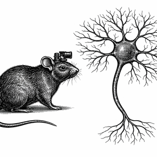
That is where this study gets interesting. The researchers looked at direct-pathway spiny projection neurons, or dSPNs, in the dorsolateral striatum of mice while the animals moved around an open field. They used calcium imaging with head-mounted miniscopes, which is a very elegant way of saying scientists strapped tiny movie cameras onto mouse brains and watched neuronal glow-sticks flicker during movement.
More Firing? Not Exactly
Here is the twist. The team found that dSPNs naturally show spatial clustering - nearby neurons tend to be co-active, especially around movement onset. But when the researchers changed signaling through mGluR5, a receptor known to shape corticostriatal synapses, they changed movement and this local co-activity pattern without much changing the overall level of dSPN activity [1].
That matters. A lot.
Blocking mGluR5 with fenobam made mice spend more time resting and shortened their movement bouts. At the same time, nearby dSPNs became more co-active. Pushing mGluR5 the other way with a positive modulator had the opposite flavor: less time resting and less local co-activity. So the dial was not "make neurons louder." It was closer to "make these neurons less likely to all yell over each other at once" [1].
If your mental image of brain function is a stadium crowd doing the wave, this paper says the wave itself matters - not just how many people are in the stadium.
Why That Is Sneakily Important
A lot of neuroscience has treated firing rate as the headline stat. Fair enough. But this study argues that the pattern of co-activation across neighboring neurons can carry its own behavioral weight. Same number of active cells, different social arrangement, different motor output.
The authors also deleted mGluR5 specifically in dSPNs, and the mice again showed reduced spontaneous movement plus more clustered dSPN co-activity. That helps pin the effect to the striatum itself instead of some whole-brain side quest. They also saw altered synaptic properties at corticostriatal inputs, which fits the idea that synaptic tuning helps set these network patterns in the first place [1].
This lines up with a broader shift in the field. Recent work argues the basal ganglia are not a simple gas pedal versus brake pedal system. Reviews in Neuron, Nature Neuroscience, Current Opinion in Neurobiology, and Trends in Neurosciences all point toward richer cell-type-specific plasticity and more nuanced direct-indirect pathway coordination than the old textbook slogan suggests [2-4,6].
Why You Should Care Even If You Do Not Own a Microscope
Movement disorders like Parkinson's disease are not just "not enough movement" problems. They are patterning problems. Timing problems. Circuit-state problems. That is why symptoms like freezing of gait can feel so bizarre and stubborn - the hardware is there, but the neural traffic system starts acting like four malfunctioning stoplights at one intersection.
So if this kind of mechanism holds up across more studies, it hints at a better therapeutic logic. Instead of only asking how to boost or suppress activity, researchers might ask how to restore healthier coordination among specific striatal ensembles. That could matter for drug development, circuit-targeted therapies, and even how we think about deep brain stimulation. Not a cure tomorrow. Not a miracle mouse-to-human leap. But a sharper map.
The big takeaway is deliciously annoying: your motor system may work best not when its neurons do more, but when they do less together in exactly the right way. The brain really is that person who says, "I do not mind if you talk, just not all at once."
References
- Marshall JJ, Xu J, Yeh NH, Yun S, Nomura T, Armstrong JN, Parker J, Contractor A. Synaptic mechanisms modulate the spatiotemporal dynamics of striatal direct pathway neurons and motor output. eLife. 2026;13:RP98122. DOI: https://doi.org/10.7554/eLife.98122. PMCID: https://pmc.ncbi.nlm.nih.gov/articles/PMC12844889/
- Roth RH, Ding JB. Cortico-basal ganglia plasticity in motor learning. Neuron. 2024;112(15):2486-2502. DOI: https://doi.org/10.1016/j.neuron.2024.06.014. PMCID: https://pmc.ncbi.nlm.nih.gov/articles/PMC11309896/
- Fang L, Creed M. Updating the striatal-pallidal wiring diagram. Nature Neuroscience. 2024;27(1):15-27. DOI: https://doi.org/10.1038/s41593-023-01518-x. PMCID: https://pmc.ncbi.nlm.nih.gov/articles/PMC11892008/
- Gomez-Ocadiz R, Silberberg G. Corticostriatal pathways for bilateral sensorimotor functions. Current Opinion in Neurobiology. 2023;83:102781. DOI: https://doi.org/10.1016/j.conb.2023.102781.
- Varin C, Cornil A, Houtteman D, Bonnavion P, de Kerchove d'Exaerde A. The respective activation and silencing of striatal direct and indirect pathway neurons support behavior encoding. Nature Communications. 2023;14(1):4982. DOI: https://doi.org/10.1038/s41467-023-40677-0. PMCID: https://pmc.ncbi.nlm.nih.gov/articles/PMC10435545/
- Varin C, de Kerchove d'Exaerde A. Neuronal encoding of behaviors and instrumental learning in the dorsal striatum. Trends in Neurosciences. 2025;48(1):77-91. DOI: https://doi.org/10.1016/j.tins.2024.11.003.
Disclaimer: The image accompanying this article is for illustrative purposes only and does not depict actual experimental results, data, or biological mechanisms.