
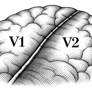
This is going to sound strange, but the researchers seem to have had a genuine what-on-earth moment when their own data kept pointing to the same odd place: the border between primary visual cortex and secondary visual cortex. Not the whole cortex. A border. In this study, the primary visual cortex, or V1, carried 30%-70% more detectable somatic mutations than neighboring V2, hinting that the cells on either side may split their family tree later, and more sharply, than anyone expected (Viswanadham et al., 2025). Your cortex, apparently, contains neighborhoods with stronger opinions than some city councils.
The Brain's Secret Genealogist
The trick in this paper is both elegant and slightly ridiculous in the best way. The authors used somatic mutations as natural barcodes. These are DNA typos that pop up after conception, then get passed to daughter cells. If two cells share the same rare mutation, they probably came from a common ancestor cell.
That lets scientists reconstruct lineage in the human brain after the fact. No fluorescent labels, no time machine, just the accumulated scribbles in the genome. Earlier work showed this approach can reveal a deeply mixed, mosaic brain rather than a neatly sorted one (Bizzotto and Walsh, 2022). This new paper pushes that idea harder with deep genome sequencing, region-by-region sampling, and single-cell transcriptomics across more than 72,000 cells.
Not All Cortex Plays By The Same Rules
One headline finding is that the fronto-parietal cortex seems comparatively relaxed about clone mixing. Mutation-marked clones there disperse broadly into neighboring regions, which fits an early developmental scramble in which related cells spread out like concertgoers who all swore they would stay together and then absolutely did not.
The visual cortex was different. Around the V1-V2 border, clones looked more spatially restricted. You might expect neighboring regions to shade gently into each other, like watercolor. Instead, this paper suggests that at least some of their cellular lineages behave more like a sharp pencil line.
That sharpness echoes another recent study, which used spatial transcriptomics to show an abrupt molecular transition between fetal V1 and V2 by mid-gestation, rather than just a smooth gradient across the cortex (Qian et al., 2025). So the new result does not arrive out of nowhere.
The Cell Family Drama Gets Better
The paper also gets into a long-running developmental argument: how separate are excitatory and inhibitory neuron lineages in the human cortex? In humans, clean stories have a way of showing up overdressed and leaving early.
Using single-nucleus mutation detection plus transcriptomes, the authors found glutamatergic neuron clones that shared low-level somatic mutations with some GABAergic neurons. Translation: some cells with different jobs may still have a surprisingly recent common ancestor. That lines up with other recent human work suggesting that at least some inhibitory neurons may share a dorsal cortical origin with excitatory populations (Chung et al., 2024, PMCID: PMC11194162).
This is where the paper gets genuinely interesting. It suggests that cortical regions are shaped not only by gene expression programs or incoming connections, but also by who is related to whom, when those branches split, and how far the descendants wander. Development, in other words, is genealogy with a migration problem.
Why You Should Care, Even If You Did Not Wake Up Wanting Clonal Architecture
A better map of human cortical lineage helps answer a basic question: how does the cortex become different from place to place while still being built from shared starting material? Recent epigenomic work already showed that human cortical progenitors vary more by region than older simple models allowed (Ziffra et al., 2021). This paper adds a lineage layer to that story.
That matters for disease. Somatic mutations are already implicated in cortical malformations, epilepsy, and some neurodevelopmental disorders (Bizzotto and Walsh, 2022). If normal human cortex contains region-specific lineage rules, then pathogenic mosaic mutations may as well. A mutation landing early in a widely dispersing clone could produce a very different pattern from one landing later near V1-V2.
There is also a quieter implication. Brain maps from imaging, transcriptomics, and anatomy often treat cortical areas as if their borders are just there, like lines on a finished painting. Studies of arealization increasingly suggest those borders emerge through layered developmental processes (Petersen et al., 2024). This paper gives us one more brushstroke in that picture: lineage itself may help draw the edges.
Which is a very brain thing to do. Even when you zoom down to tiny mutations in single cells, the cortex still refuses to be boring. It does not build itself like a factory line. It builds itself like an overcomplicated fresco - sketched, revised, crowded, and occasionally divided by a line so crisp the artists themselves had to stop and stare.
References
- Viswanadham VV, Kim SN, Caglayan E, et al. Combined somatic mutation and transcriptome analysis reveals region-specific differences in clonal architecture in human cortex. Cell Reports. 2025;44(11):116458. https://doi.org/10.1016/j.celrep.2025.116458
- Bizzotto S, Walsh CA. Genetic mosaicism in the human brain: from lineage tracing to neuropsychiatric disorders. Nature Reviews Neuroscience. 2022;23:275-286. https://doi.org/10.1038/s41583-022-00572-x
- Ziffra RS, Kim CN, Ross JM, et al. Single-cell epigenomics reveals mechanisms of human cortical development. Nature. 2021;598:205-213. https://doi.org/10.1038/s41586-021-03209-8
- Chung C, Yang X, Hevner RF, et al. Cell-type-resolved mosaicism reveals clonal dynamics of the human forebrain. Nature. 2024;629:384-392. https://doi.org/10.1038/s41586-024-07292-5. PMCID: https://pmc.ncbi.nlm.nih.gov/articles/PMC11194162/
- Qian X, Coleman K, Jiang S, et al. Spatial transcriptomics reveals human cortical layer and area specification. Nature. 2025;644:153-163. https://doi.org/10.1038/s41586-025-09010-1
- Petersen SE, Seitzman BA, Nelson SM, Wig GS, Gordon EM. Principles of cortical areas and their implications for neuroimaging. Neuron. 2024;112(17):2837-2853. https://doi.org/10.1016/j.neuron.2024.05.008
Disclaimer: The image accompanying this article is for illustrative purposes only and does not depict actual experimental results, data, or biological mechanisms.