The last time you reached for your phone, your brain was secretly running a civic disaster of astonishing competence; millions of neurons exchanged tiny electrical rumors, inhibitory cells played bouncer, and your cortex somehow turned "maybe check one message" into ten minutes of doomscrolling. That ordinary act is the sort of thing neuroscience still struggles to explain in full, because the brain is less like a neat machine and more like the Ship of Theseus after several espressos - rebuilt, layered, recursive, and held together by consequences. A new paper in eLife tackles that mess by building a giant, biophysically detailed model of rat somatosensory cortex and asking whether a simulated cortex can behave enough like the real one to teach us something worth keeping [1].
The Brain Flight Simulator Nobody Asked For, But Everybody Needed
The headline numbers are mildly absurd in the best way: eight cortical subregions, 4.2 million morphologically and electrically detailed neurons, and 13.2 billion synapses [1]. This is not a toy network with a few cartoon neurons firing in a polite spreadsheet. It is an attempt to model microcircuitry and mesocircuitry together - local wiring plus the mid-range links between nearby cortical areas - so researchers can test ideas that would be painfully difficult, expensive, or flat-out impossible to probe in a living brain. Real cortex does not reveal its secrets on demand; you can record from some cells and stimulate others, but the computation emerges from many scales at once.
When a Simulation Has to Earn Your Trust
The impressive part is not that the model is huge; big things are easy to make if you are willing to build a cathedral of nonsense. The impressive part is that it reproduced several experimental findings under a single parameterization, including millisecond-precise responses to sensory stimuli, patterned responses under targeted optogenetic activation, and selective spread of activity to downstream regions [1]. In plain English: the fake cortex did not just sit there looking expensive. It behaved enough like laboratory cortex to earn the right to be interrogated.
The Interneurons Are Not "Support Staff"
One of the paper's most interesting angles involves inhibitory interneurons - the cells that stop cortex from turning into a screaming electrical mosh pit. The model incorporated more specific targeting rules for inhibitory subpopulations, including rules informed by electron microscopy work showing that different inhibitory cell types do not distribute their synapses randomly [1]. That fits a larger 2025 connectomics wave: a Nature study mapped inhibitory specificity in mouse visual cortex and found structured motif-like targeting rather than generic "slow everybody down" behavior [2]. We often talk about excitation as the hero and inhibition as the stern librarian; but the librarian is also editing the plot. The model predicts that different inhibitory cell types and cortical layers contribute differently to stimulus encoding [1].
Why This Weird Monster Could Matter Outside the Server Room
If these results keep holding up, models like this could become the neuroscience version of wind tunnels or flight simulators. You would not replace real experiments; you would stop wasting real animals and real time on every bad guess. Large cortical models could help researchers test how circuit motifs shape perception, predict the effects of stimulation, or narrow which cell types matter most before heading back to the lab.
The timing is not random. On April 9, 2025, the MICrONS consortium released a major functional connectomics dataset across mouse visual cortex, and companion studies tied function to detailed wiring rules [3,4]. Around the same period, experts were openly talking about "digital twins" for neuroscience - not in the sci-fi sense where your laptop develops opinions, but as adaptive models that connect anatomy, physiology, and prediction in a usable framework [5]. Even Nature Methods named EM connectomics its 2025 Method of the Year, which is a polite editorial way of saying: the wiring-diagram era has arrived and is now rearranging the furniture.
There are still limits. A model this broad is hard to validate everywhere at once, and some claims rest on smaller subvolumes or specific manipulations rather than the full cortex-scale beast [1]. But that is the honest state of the field. Nobody has built Plato's ideal cortex in silicon. What we have instead is an increasingly testable argument about how real cortical structure gives rise to real cortical dynamics. The cortex still feels like a fog that somehow writes symphonies and loses arguments to late-night snacks; this paper does not clear the fog, but it gives it edges.
References
- Isbister JB, Ecker A, Pokorny C, et al. Modeling and simulation of neocortical micro- and mesocircuitry (Part II, Physiology and experimentation). eLife. 2026;13:RP99693. DOI: https://doi.org/10.7554/eLife.99693.3. PubMed: https://pubmed.ncbi.nlm.nih.gov/41556778/
- Schneider-Mizell CM, Bodor AL, Brittain D, et al. Inhibitory specificity from a connectomic census of mouse visual cortex. Nature. 2025;640(8058):448-458. DOI: https://doi.org/10.1038/s41586-024-07780-8. PubMed: https://pubmed.ncbi.nlm.nih.gov/40205209/ PMCID: https://pmc.ncbi.nlm.nih.gov/articles/PMC11981935/
- MICrONS Consortium. Functional connectomics spanning multiple areas of mouse visual cortex. Nature. 2025;640(8058):435-447. DOI: https://doi.org/10.1038/s41586-025-08790-w. PubMed: https://pubmed.ncbi.nlm.nih.gov/40205214/ PMCID: https://pmc.ncbi.nlm.nih.gov/articles/PMC11981939/
- Ding Z, Fahey PG, Papadopoulos S, et al. Functional connectomics reveals general wiring rule in mouse visual cortex. Nature. 2025;640(8058):459-469. DOI: https://doi.org/10.1038/s41586-025-08840-3. PubMed: https://pubmed.ncbi.nlm.nih.gov/40205211/
- Fekonja LS, Schenk R, Schröder E, et al. The digital twin in neuroscience: from theory to tailored therapy. Front Neurosci. 2024;18:1454856. DOI: https://doi.org/10.3389/fnins.2024.1454856. PubMed: https://pubmed.ncbi.nlm.nih.gov/39376542/ PMCID: https://pmc.ncbi.nlm.nih.gov/articles/PMC11457707/
- Senk J, Kriener B, Djurfeldt M, et al. Connectivity concepts in neuronal network modeling. PLoS Comput Biol. 2022;18(9):e1010086. DOI: https://doi.org/10.1371/journal.pcbi.1010086. PubMed: https://pubmed.ncbi.nlm.nih.gov/36074778/ PMCID: https://pmc.ncbi.nlm.nih.gov/articles/PMC9479696/
- Hanganu-Opatz IL, Butt SJB, Hippenmeyer S, et al. The Logic of Developing Neocortical Circuits in Health and Disease. J Neurosci. 2021;41(5):813-822. DOI: https://doi.org/10.1523/JNEUROSCI.1655-20.2020. PubMed: https://pubmed.ncbi.nlm.nih.gov/33431633/
- Nandi A, Chartrand T, Van Geit W, et al. Single-neuron models linking electrophysiology, morphology, and transcriptomics across cortical cell types. Cell Rep. 2022;40(6):111176. DOI: https://doi.org/10.1016/j.celrep.2022.111176. PubMed: https://pubmed.ncbi.nlm.nih.gov/35947954/ PMCID: https://pmc.ncbi.nlm.nih.gov/articles/PMC9793758/
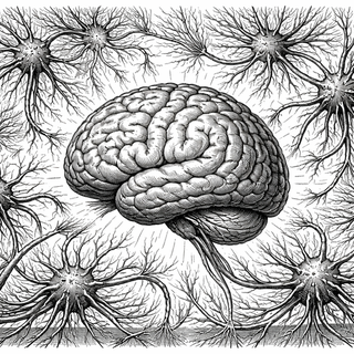
Disclaimer: The image accompanying this article is for illustrative purposes only and does not depict actual experimental results, data, or biological mechanisms.